
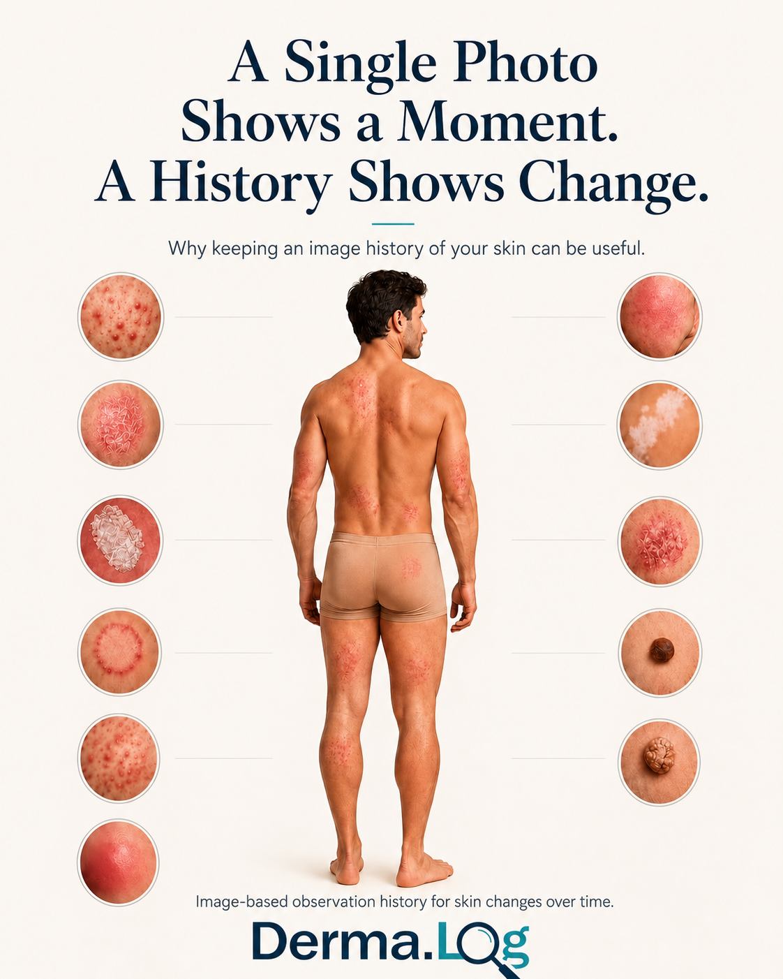
Why image history matters
Skin can look very different from one person to another—and from one point in time to another.
A mark may disappear within days. Another may remain almost unchanged for years. Some changes happen gradually enough that remembering exactly what an area looked like three, six, or twelve months ago can be surprisingly difficult.
A photograph gives you something memory cannot: a reference. When photographs are kept in sequence, they can become something more useful than isolated pictures. They become a history.
Many leading dermatology and cancer organizations recommend recording or photographing skin changes over time.
The American Academy of Dermatology explains that photographs can help a dermatologist see how a rash, growth, or other skin concern is changing. The British Association of Dermatologists says that a photograph can be helpful when monitoring a mole for change. Cancer Council Australia similarly recommends photographing moles and spots so newer images can be compared with older ones.
Cancer Research UK describes photography as part of clinical observation because photographs make changes over time easier to review. The Skin Cancer Foundation recommends documenting findings during skin self-examination, including by taking pictures. In guidance for people after melanoma treatment, Denmark's Patienthåndbogen also discusses self-observation and notes that many people use mobile-phone photographs to help follow their skin.
These organizations support the principle of recording and comparing change; they do not endorse Derma.Log. The shared idea is not that a photograph can tell you what something is. It is that a previous photograph gives you something concrete to compare with.
Why memory isn't enough
You may remember that a Spot was present. You may remember approximately how large or red it seemed. More precise questions are harder to answer from memory alone:
- Was that edge already shaped like this?
- Was the color the same?
- Was there scaling last time?
- Did the redness extend this far?
- Has the appearance actually changed?
- When did I first notice it?
Looking at something frequently can make gradual change especially difficult to perceive. A previous image gives you a reference point. A sequence of images gives you chronology.
Taking useful comparison photographs
If images are going to be compared later, image quality and context matter. The American Academy of Dermatology recommends several simple principles:
- Use good, even lighting.
- Avoid filters.
- Make sure the image is in focus.
- Use a neutral background where possible.
- Show what the skin actually looks like.
- Take more than one perspective when useful.
Its photography guidance describes three particularly useful perspectives:
- An orientation photograph showing where the area is located.
- A comparison photograph showing the area together with surrounding skin.
- A close-up showing the visible detail.
A ruler or another familiar size reference can sometimes help provide scale when appropriate. The purpose is not to make a beautiful photograph. The purpose is to preserve an accurate visual reference.
Photographs cannot diagnose
A photograph can preserve appearance. It cannot reproduce everything a clinician can assess. Texture, palpation, symptoms, clinical history, dermoscopy, and other examination findings may all matter.
Cancer Research UK describes photography as a way to make change easier to review over time—not as a replacement for professional assessment. The Skin Cancer Foundation makes the same distinction: photographs can be useful reference material, while professional skin examination remains important.
That distinction is fundamental to Derma.Log. Derma.Log is an observation and record-keeping tool. It is not a diagnostic scanner, does not detect or rule out cancer, and does not replace a qualified medical professional. The site's Medical Disclaimer explains these boundaries in more detail.
The camera roll problem
Taking a skin photograph is easy. Finding and understanding it six months later can be much harder. It may sit somewhere between holiday photographs, screenshots, receipts, family photographs, pets, food, work images, and thousands of other pictures.
Even when you find the photograph, the context may be gone. Which area was this? When was it taken? Was this before or after the other photograph? What had you noticed at the time? Were you planning to look at it again?
A camera roll stores photographs. It does not organize the story behind them. Skin photographs can also be sensitive personal data, so consider where they are stored and how they are shared. The Derma.Log Privacy Policy explains how app records, exports, and external copies are handled.
How Derma.Log organizes image history
Derma.Log is built around one simple relationship:
Spot Log Image
A Spot represents one particular skin area or observation you want to follow. Each time you return to that Spot, you can create another Log. That Log represents one observation point in time. One or more images belonging to that Log stay attached to that observation instead of becoming disconnected pictures.
Over time, those Logs form the history of that Spot. Instead of asking, “Where is that old photo?” you can ask, “What did this same Spot look like before?”
This organization supports documentation and review. It does not decide what an image means or make a medical judgment. If you need help using the app, Derma.Log Support can assist with product questions but cannot provide medical advice.
Images first. Context alongside them.
The image remains the primary visual reference. Derma.Log also lets the user record structured observations alongside an individual Log. The current observation signals are:
- Inflammation
- Oozing
- Scaling
- Color Change
- Border Change
These signals belong to the observation at that point in time. They do not replace the photograph or provide a diagnosis. They add context to the record so the image and what you recorded alongside it can be reviewed together later.
Come back when it makes sense
There is no universal image-taking interval that is right for every person or every skin concern. The American Academy of Dermatology recommends asking your dermatologist how often photographs should be taken when photography is being used to follow a concern.
Derma.Log does not decide a medical follow-up schedule. Its reminders are organizational tools. You decide what you want to return to and when; a reminder is not medical guidance.
Take the history with you
Sometimes a history remains useful only to you. Sometimes you may later want to discuss what you observed with a healthcare professional. There is a practical difference between saying, “I think it changed,” and being able to show an earlier photograph, later observations, and their sequence.
Derma.Log includes structured export tools so recorded history can be prepared outside the app when needed. Review exported material before sharing it and choose a trusted destination. An export is not a clinician-created medical record unless a clinician separately reviews and adopts it. The app does not make the medical judgment; its purpose is to help preserve the information.
A simple Derma.Log workflow
For an area of skin that you want to follow:
- Create a Spot.
- Take clear photographs of what you actually see.
- Record the observation.
- Add another Log when you return to it.
- Compare the history instead of relying only on memory.
- Add a reminder if you want to revisit it later.
- Export the history if you want to take the information with you.
One photograph shows one moment. A second photograph gives you a comparison. A series of photographs gives you a history—and sometimes that history is much more useful than trying to remember.
Your skin history, organized.
Derma.Log helps keep photographs, observations, and chronology connected to the same Spot over time.